Building the biological and biomaterial foundations for urethral tissue engineering
Histological section of human tissue. Source: Envato; Micro-CT of a hare’s urethra. Source: UMC Utrech and UGent; Hydrogel being prepared for testing. Source: 4Tissue; A rheometer applies force onto hydrogel. Source: 4Tissue;
Conceptual visualisation of the 3D bioprinting approaches STRONG-UR is researching to treat male urethral strictures. Source: STRONG-UR
Urethral defects and strictures can severely affect a patient’s quality of life and remain among the most challenging conditions in reconstructive urology. Although current surgical approaches achieve good outcomes for most patients, they often rely on harvesting healthy tissue, which is not always feasible and may lead to complications. To address these limitations, researchers are exploring 3D bioprinting to create tissue-engineered grafts capable of restoring both the structure and function of damaged urethral tissue.
In response, the STRONG-UR team brings together expertise in clinical urology, tissue biology, biomaterials science, biofabrication, and regulatory science. By integrating biological understanding with material innovation from the earliest stages, STRONG-UR aims to establish both the design requirements and the technological foundations for next-generation regenerative therapies.
The first phase of the project focused on two complementary questions:
- What characteristics define a functional urethra and therefore need to be recreated?
- Which biomaterials show enough promise to support tissue regeneration of these key urethral characteristics, while demonstrating encouraging performance in early safety evaluations?
This work led to the successful completion of the project’s first two major milestones:
- Establishing a detailed biological and mechanical blueprint of the male urethra.
- Identifying promising hydrogel formulations for continued bioprinting development.
Together, these milestones represent important early steps toward the long-term goal of creating safe, functional, and patient-specific regenerative solutions for urethral reconstruction.
Understanding the male urethra in health and disease
To rebuild a tissue, we first need to understand what it is made of and how it works. For the male urethra, this means looking closely at the layers, structures, and mechanical properties that allow it to function.
The STRONG-UR team set out to build a detailed picture of the urethra, establishing a biological and mechanical blueprint and setting the benchmarks that future bioprinted substitutes must meet.
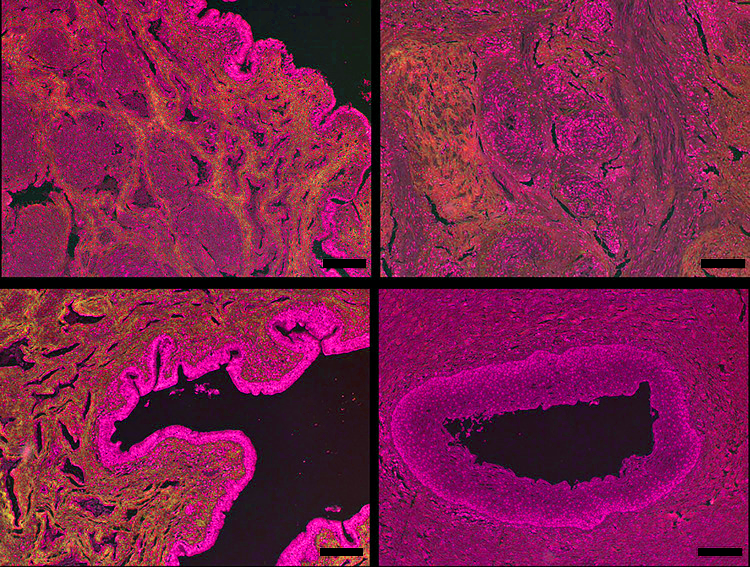
Collagen organisation in healthy (left) vs. diseased (right) urethral tissue. Source: Linssen et al. (2023). Extracellular matrix analysis of fibrosis: A step towards tissue engineering for urethral stricture disease. PloS one, 18(11), e0294955. https://doi.org/10.1371/journal.pone.0294955
Collagen organisation in healthy (left) vs. diseased (right) urethral tissue. Source: Linssen et al. (2023). Extracellular matrix analysis of fibrosis: A step towards tissue engineering for urethral stricture disease. PloS one, 18(11), e0294955. https://doi.org/10.1371/journal.pone.0294955
3D-reconstruction of the urethral surface and the spongy tissue. Source: UMC Utrecht
It is tempting to view the urethra as a simple conduit whose function is merely to convey urine. But, in reality, the male urethra is a complex, living structure composed of specialised, nested layers. It is anatomically divided into the prostatic, membranous, and spongy segments. The spongy (anterior) portion accounts for 70–85% of its length and is the most common site of stricture formation.
The urethra is also mechanically active. Most of the time it is closed, and it opens like a valve to convey urine or semen. During voiding or erection, it is compressed, stretched, or expanded. Its structure must cope with all these mechanical demands.
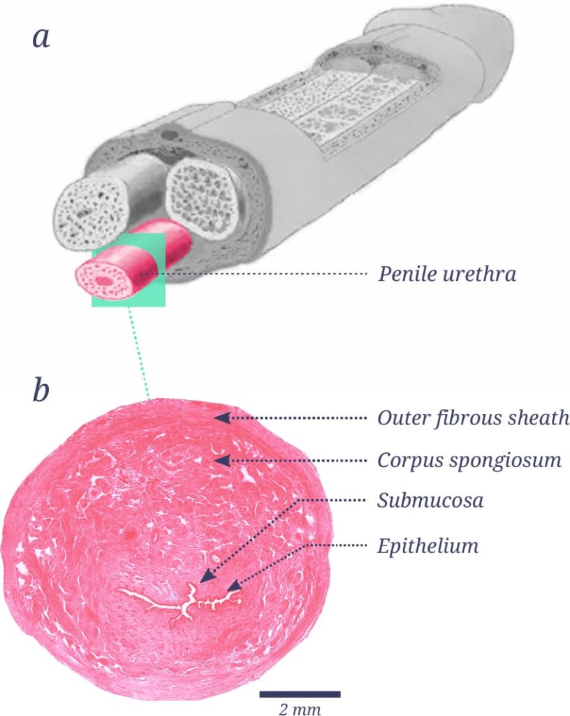
a. Penile anatomy highlighting the penile urethra. Source: this image is reproduced and adapted from Calogero et al. (2018). https://doi.org/10.1080/13685538.2018.1430758.
b. Constituent layers of the human male spongy urethra shown on a histological transverse section. Source: UMC Utrecht
Moving from the inside out, each layer of the spongy urethra performs a distinct role:
- The epithelium: The innermost lining is regionally specialised. Near the external opening, it acts as a robust protective barrier against mechanical friction. More proximally, it transitions into a lining associated with mucus-secreting glands, which lubricates the channel and shields the underlying tissue from the chemical irritation of urine.
- The submucosa: Lying directly beneath the epithelium, this dense inner layer is enriched in collagen and elastin fibres and has an abundant network of small blood vessels.
- The corpus spongiosum: Surrounding the submucosa, this soft, highly vascularised and porous tissue provides both mechanical support and the principal blood supply that sustains the urethra.
- The outer fibrous sheath: Enclosing the organ, this tough, fibrous layer helps distribute physical pressure evenly and prevents the tissue from stretching too much.
Clinically, superficial injury confined to the epithelium and submucosa does not necessarily result in urethral stricture. However, when damage extends into the corpus spongiosum, fibrotic remodelling can compromise local blood supply and tissue compliance, facilitating stricture progression and increasing the risk of recurrence after surgery.
Together, these layers illustrate a fundamental biological principle: the structure–function relationship. How the urethra is physically built is precisely what enables it to perform its physiological roles, and altering the structure alters what the tissue can do. Understanding this relationship allows STRONG-UR to design bioprinted grafts that aim to reproduce the native mechanics, barrier function, and tissue architecture of the healthy urethra.
Collagen organisation in healthy (left) vs. diseased (right) urethral tissue, highlighting the structural remodeling and increased stiffness engineered grafts must withstand. Source: Linssen et al. (2023). Extracellular matrix analysis of fibrosis: A step towards tissue engineering for urethral stricture disease. PloS one, 18(11), e0294955. https://doi.org/10.1371/journal.pone.0294955
Why study both healthy and diseased tissue?
To design a successful graft, studying healthy tissue alone is not enough. Healthy urethral tissue is regulated by many factors, including age, hormones, and blood supply and metabolic status. For example, ageing is associated with increased collagen content and altered elastic fibres, while hormonal changes such as androgen suppression can disturb the balance between the lining and the corpus spongiosum. These subtle changes in composition influence how the tissue behaves mechanically.
Disease, however, does not merely damage the urethra; it fundamentally remodels it. When trauma or medical procedures disrupt the protective epithelial lining, urine leaks into the underlying layers and causes inflammation, eventually leading to fibrosis (scarring) and stricture. As a result, diseased tissue becomes stiffer than healthy tissue, its blood supply becomes compromised, and it no longer responds mechanically as it should, impeding urination.
Examining diseased tissue alongside healthy tissue serves two purposes for STRONG-UR:
- Preparing for the implant environment: A graft designed only on the basis of healthy tissue would risk being placed in an environment it was never built to withstand and could fail for reasons that had not been anticipated. Understanding the pathological changes shows what any engineered replacement must be able to endure.
- Building better pre-clinical models: Mapping disease progression will help the consortium create better laboratory models for catheter testing and disease modelling.
Biomechanical properties
- Stiffness: how strongly the tissue resists stretching
- Elasticity: how well it stretches and springs back.
Techniques range from stretching a whole tissue segment to gently pressing on individual layers (nanoindentation) to see how firm each one is. Because scarring gradually stiffens the urethra and robs it of its natural flexibility, measuring stiffness also gives a useful marker of how far the disease has progressed.
These measurements establish clear target benchmarks for our hydrogels: engineered grafts need to be soft enough to stretch open during urination, yet strong enough to hold surgical stitches and withstand internal pressure.
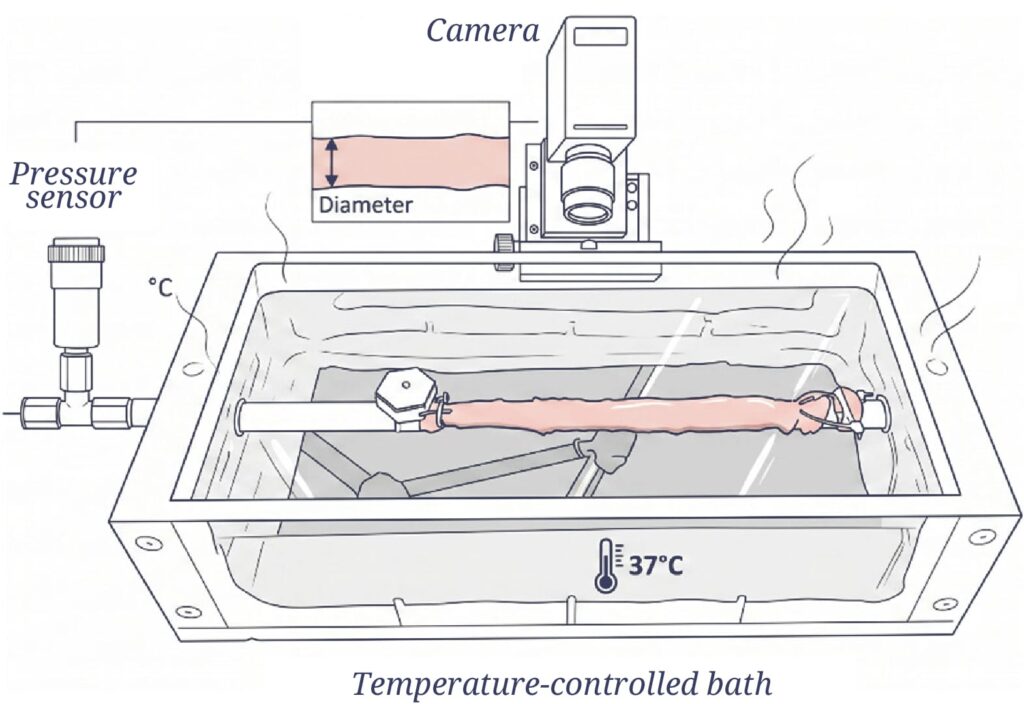
Schematic of an experimental device developed to record the intraluminal pressure-diameter relationship in urethral tissues. It consists of a physiological heated bath containing the urethra mounted on tubular rods, a pressure sensor, a syringe pump to perfuse liquid, and a camera above to record diameter changes. Original figure incorporating artwork from SciDraw by I.Martinier, CC BY 4.0.
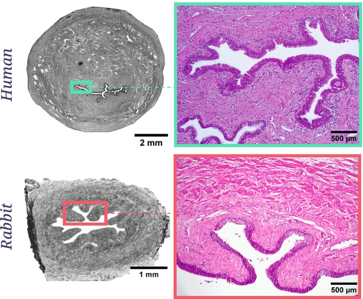
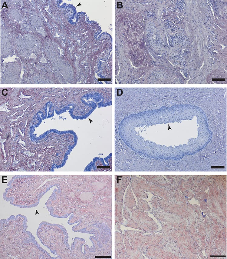
Transversal sections of human and rabbit urethral tissues and a magnified view of their luminal epithelium. Source: UMC Utrecht
Why the rabbit model?
Translating these biological blueprints into real-world therapies requires a reliable pre-clinical animal model. However, no consensus has been reached on which species best resembles human penile anatomy, and the animals used in urology vary widely, some with anatomy far from that of the human.
Following a literature review, the rabbit consistently emerged as the best anatomical match for the human male urethra. Its epithelial lining, lumen diameter, and, crucially, its surrounding corpus spongiosum closely resemble the human counterpart. Because the corpus spongiosum provides structural support and drives wound healing, a faithful model of urethral tissue engineering needs an animal with similar tissue around the urethra. Weighing anatomy, disease modelling, and surgical handling against cost and ethical considerations, the rabbit offers the most favourable combination for translational work, making it a suitable platform to test new graft materials before moving towards the clinic.
A blueprint for bioprinted substitute tissue
We cannot rebuild a tissue we have not first understood. By establishing a biological and mechanical blueprint of both healthy and diseased urethral tissue, this milestone sets performance benchmarks that any bioprinted substitute must meet, and translates clinical needs into concrete mechanical and biological targets for hydrogel development.
With this blueprint in hand, the next step is to identify materials that could recreate it and to check that they show encouraging early performance.
Identifying biomaterials for future regenerative therapies
Once the biological and mechanical features of the male urethra were established, the next challenge was to identify biomaterials capable of recreating them.
An ideal material must support living cells while possessing physical properties suited to bioprinting. To meet these demands, the STRONG-UR team evaluated a series of hydrogel-based formulations.
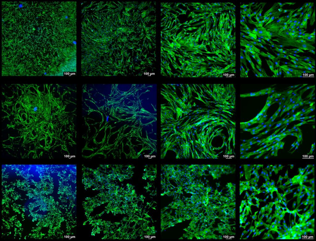
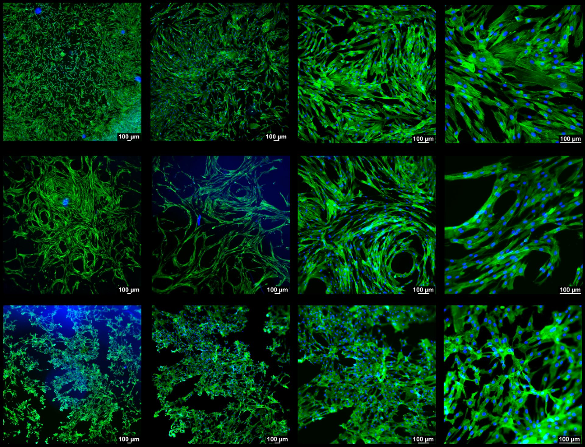
2D monoculture of human adipose stem cells (hASCs) in hydrogel at various magnifications. Source: Tampere University
2D monoculture of human adipose stem cells (hASCs) in hydrogel at various magnifications. Source: Tampere University
Preparation of hydrogel to test its mechanical properties. Source: 4Tissue
What are hydrogels?
Hydrogels are water-rich materials widely used in regenerative medicine because their soft, hydrated nature mimics aspects of the natural environment around cells. They can be engineered with different stiffness levels and biological properties, making them versatile building blocks for bioprinting soft tissues such as the urethra.
STRONG-UR focuses on hydrogels based on two complementary building blocks:
- Gelatine: A protein derived from collagen, a main structural component of the body’s tissues. Gelatine provides natural biological signals that help cells attach, grow, and organise into tissue.
- Dextran: A natural sugar-based polymer (polysaccharide) that can mimic some functions of glycosaminoglycans (GAGs), molecules in native tissue that contribute to hydration and mechanical behaviour.
By combining gelatine and dextran, STRONG-UR aims to create biomaterials that reproduce key aspects of the native cellular environment while providing the physical properties needed for bioprinting. These materials can be tailored to the biological and mechanical targets identified in the first milestone.
Evaluating promising biomaterial candidates
Not all hydrogels are equally suitable for regenerative applications. Our researchers investigated both single-component hydrogels (based on gelatine or dextran) and hybrid formulations combining the two materials in different proportions. Selected formulations were subjected to a series of laboratory-based evaluations of both their biological performance and their physical characteristics.
These early in vitro assessments provide an opportunity to identify potential risks before moving to more complex models. To guide this process, the consortium applied testing approaches based on ISO 10993 standards, an internationally recognised framework for the biological evaluation of materials intended for medical applications. This supports a systematic and transparent evaluation, including checks for harmful effects on cells.
Specifically, the team investigated three key questions:
- Cellular support: Can the materials support cell survival and normal cellular activity?
- Mechanical compatibility: Do they have physical properties compatible with the mechanical demands of urethral tissue?
- Stability: Do they remain sufficiently stable under laboratory conditions that mimic the physiological environment?
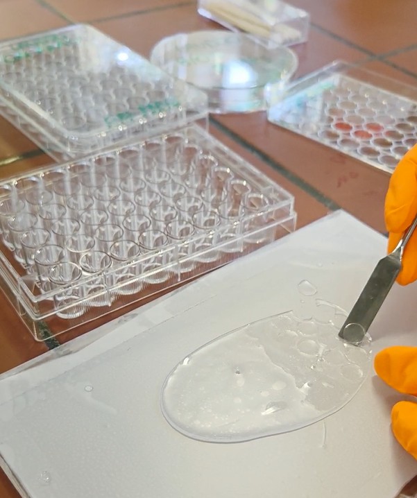
Preparation of a hydrogel swellability/swelling test. Source: 4Tissue
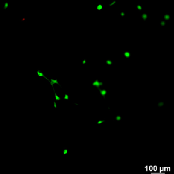
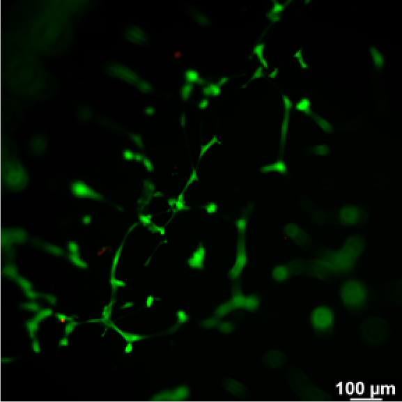
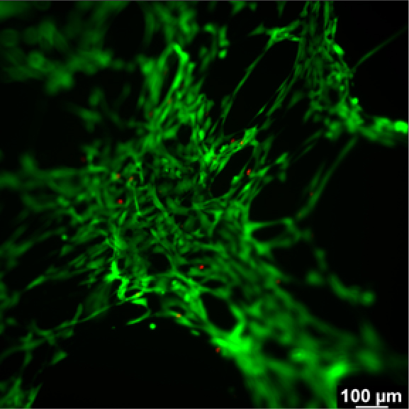
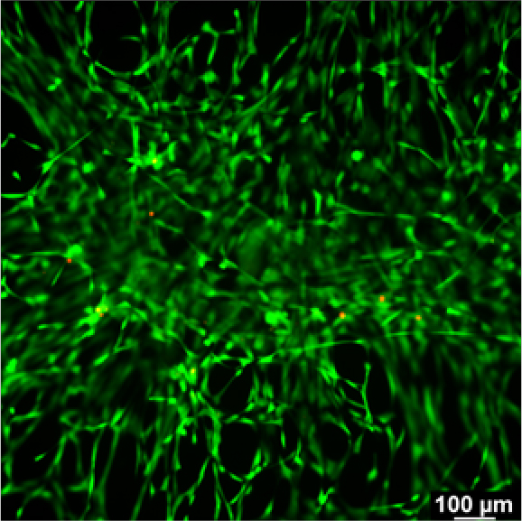
2D monoculture of human adipose stem cells (hASCs) in hydrogel over 14 days. Source: Tampere University
What did we learn?
The evaluated hydrogel formulations demonstrated encouraging performance in these initial studies. The selected materials supported cell viability and showed no evidence of cytotoxic (cell-damaging) effects under the tested conditions. The tests also provided valuable insights into how material composition influences cell growth, helping narrow the field to the most promising formulations for further optimisation.
It is important to note that these laboratory evaluations are an early step in a much broader development pathway. Showing that a material causes no detectable cytotoxic effects in the laboratory does not establish overall biocompatibility or clinical safety. Instead, these studies help identify promising candidates and focus future biofabrication efforts on the materials most likely to meet the biological and mechanical targets established in the first milestone.
Impact and the future of regenerative medicine
The achievements of these milestones are not separate accomplishments, but complementary steps within a single pathway. Together, they address two fundamental requirements for any future regenerative therapy: understanding what needs to be rebuilt (the biological blueprint) and identifying materials capable of supporting that process.
By combining tissue characterisation with early material assessment, STRONG-UR reduces uncertainty at an early stage and helps de-risk the development of future regenerative therapies. Applying internationally recognised ISO testing approaches also demonstrates a commitment to developing these biomaterials in a rigorous and responsible manner. The studies conducted so far represent only an early stage of the evaluation pathway required for medical applications. They do not demonstrate complete safety or clinical readiness, but they help build confidence that the selected materials and design strategies are progressing in a promising direction.
Beyond urethral reconstruction, this integrated framework may prove valuable for other tissue-engineering applications. It shows how combining detailed biological understanding with systematic material design can guide regenerative medicine research, particularly where success depends on both reproducing tissue function and choosing materials that can support it.
Looking ahead, the team will focus on refining hydrogel formulations, advancing biofabrication strategies, and evaluating performance in increasingly complex biological models. Significant challenges remain before clinical implementation can be considered, but the progress achieved so far provides a strong foundation. By grounding material innovation in true tissue biology, STRONG-UR contributes to moving the field one step closer to personalised, patient-specific regenerative solutions.
STRONG-UR scientists at work (STRONG-UR, Aalborg University), hydrogel synthesisation (STRONG-UR), extrusion-based printing (Aalborg University), and DLP printing (University of Ghent).
Article contributors
Cristian Pablo Pennisi (Aalborg University); Petra de Graaf, Isabelle Martinier (UMC Utrecht); Nele Eeckman, Lana Van Damme, (4Tissue); Nele Pien (Ghent University); Elizaveta Kuzmina, Raul Jimenez van Hoorn (META Group)